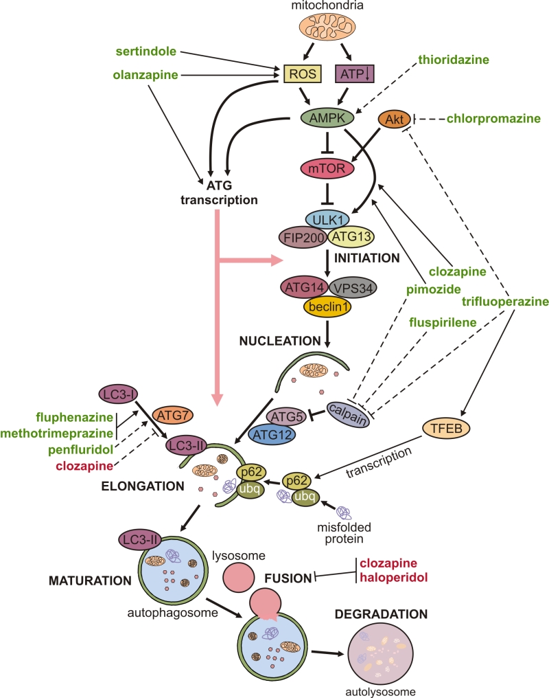

Only those cellular proteins known to be adaptors for targeting microbes are shown other proteins (not shown) also function in cargo recognition of mitochondria and other organelles. The cellular events that occur during autophagy are depicted in the bottom diagram, including the major known cellular and microbial proteins that regulate autophagy initiation, cargo recognition and autophagosome maturation. Autophagy is a natural cellular mechanism by which the cells in our body degrade unnecessary or damaged components within the cell. WIPIs and the ATG12–ATG5–ATG16L1 complex are present on the outer membrane, and LC3–PE is present on both the outer and inner membrane of the isolation membrane, which may emerge from subdomains of the ER termed omegasomes. After induction of autophagy, the ULK1 complex (ULK1–ATG13–FIP200–ATG101) (downstream of the inhibitory mTOR signalling complex) translocates to the ER and transiently associates with VMP1, resulting in activation of the ER-localized autophagy-specific class III phosphatidylinositol-3-OH kinase (PI(3)K) complex, and the phosphatidylinositol-3-phosphate (PtdIns(3)P) formed on the ER membrane recruits DFCP1 and WIPIs. The major membrane source is thought to be the endoplasmic reticulum (ER), although several other membrane sources, such as mitochondria and the plasma or nuclear membrane, may contribute. The top right box shows a model of our current understanding of the molecular events involved in membrane initiation, elongation and completion of the autophagosome. The FEBS Journal published by John Wiley & Sons Ltd on behalf of Federation of European Biochemical Societies.Overview of the autophagy pathway. Understanding the regulation of selective autophagy receptors will provide deeper insights into the pathway and open up potential therapeutic avenues.Īutophagy oligomerisation phosphorylation receptor ubiquitination. We highlight the four most prominent protein modifications - phosphorylation, ubiquitination, acetylation and oligomerisation - that are essential for autophagy receptor recruitment, function and turnover. Its a natural process of cellular repair and cleaning. Autophagy receptors undergo post-translational and structural modifications to fulfil their role in autophagy, or upon executing their role, for their own degradation. Autophagy (pronounced ah-TAH-fah-gee) is an opportunity for your cells to take out the garbage, says Cynthia Thurlow, NP, nurse practitioner and functional nutritionist who specializes in intermittent fasting.

To distinguish between different cellular components, autophagy employs selective autophagy receptors, which will link the cargo to the autophagy machinery, thereby sequestering it in the autophagosome for its subsequent degradation in the lysosome. Autophagy also controls stem cell fate and defective autophagy is involved in many pathophysiological processes. This conserved process is essential for metabolic plasticity and tissue homeostasis and is crucial for mammalian post-mitotic cells. Autophagy (or autophagocytosis from the Ancient Greek, autphagos, meaning 'self-devouring' 1 and, ktos, meaning 'hollow') 2 is the natural, conserved degradation of the cell that removes unnecessary or dysfunctional components through a lysosome-dependent regulated mechanism. Hence, autophagy can be nonselective, where bulky portions of the cytoplasm are degraded upon stress, or a highly selective process, where preselected cellular components are degraded. Autophagy is a constitutive pathway that allows the lysosomal degradation of damaged components. Autophagy is a highly conserved catabolic process cells use to maintain their homeostasis by degrading misfolded, damaged and excessive proteins, nonfunctional organelles, foreign pathogens and other cellular components.


 0 kommentar(er)
